


Full-Arch Restorations
Fixed implant-supported teeth for patients requiring complete rehabilitation.

Four areas of treatment, connected by one principle: the surgical, biological and restorative stages must be planned as one treatment.
Fixed implant-supported teeth for patients requiring complete rehabilitation.
Replacing one missing tooth while preserving the neighbouring teeth and surrounding tissues.
Reconstructive procedures that create the correct foundation for implant treatment.
Restoring the volume, contour and stability of the gums around teeth and implants.
Full-arch treatment replaces all teeth in the upper jaw, lower jaw or both jaws with a fixed restoration supported by dental implants. It is considered when multiple teeth are missing, the remaining teeth cannot be predictably maintained, or a patient is currently dependent on removable dentures and wishes to move to a fixed solution.
The objective is not simply to place implants.
The objective is to design a restoration that functions well, respects the available biology and remains maintainable over time.
FP1 and FP3 describe how the final restoration replaces the teeth and how it relates to the patient’s remaining gum and bone tissue. Neither concept is universally better. The correct choice depends on anatomy, smile line, tissue volume, facial support, implant position, hygiene and long-term predictability.
FP1 — Natural gum tissue remains visible.
The restoration replaces the clinical crowns. Each tooth must emerge through the patient’s own soft tissue with realistic proportions and carefully controlled contours.
FP3 — Lost teeth and tissue volume are restored together.
Pink restorative material replaces missing gum and bone volume, allowing natural tooth proportions and, where needed, additional facial and lip support.
FP1 represents only a small proportion of full-arch treatment worldwide because it requires advanced surgical, prosthetic and soft-tissue expertise, precise guided implant positioning and close control of every stage.
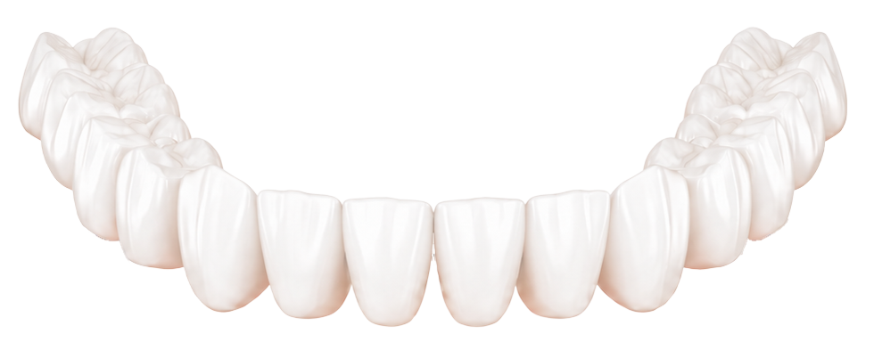

An FP1 restoration replaces the natural crown portion of the teeth. The restoration is designed to emerge through the patient’s own soft tissue, without artificial pink material.
The absence of pink ceramic alone does not define FP1. The true reference point is anatomy: the restoration must emerge realistically from the soft tissue and replace only the natural crown portion of the tooth.
FP1 is highly demanding.
When anatomy is unfavourable, trying to force an FP1 design can create over-contoured teeth, unstable tissue and a restoration that is difficult to maintain.
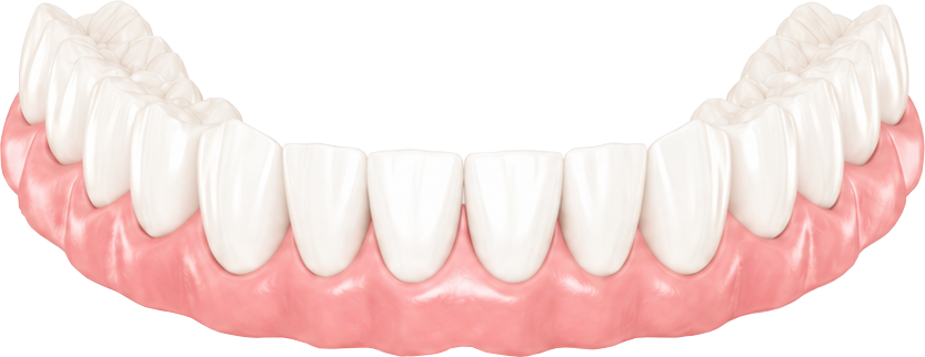
An FP3 restoration includes both white teeth and pink restorative material. It is used when the loss of bone and gum tissue would make an FP1 restoration disproportionate, unstable or surgically unrealistic.
The pink component is not simply cosmetic. It allows the restoration to recreate lost volume, restore more natural tooth proportions and, where required, provide support for the lips and lower facial profile.
FP3 is not an inferior solution.
For the right patient, it may provide the more balanced, maintainable and predictable long-term result.
We do not begin by deciding that a patient should receive FP1 or FP3. We first evaluate the clinical situation and then select the design that offers the best balance of aesthetics, function, hygiene and long-term stability.
How much tooth and gum are visible during a natural smile?
How much supporting bone remains, and where can implants be placed safely?
Is the tissue thick, stable and sufficient to create a natural emergence profile?
Can the planned teeth be created in realistic length and width?
Does the restoration need to replace lost tissue volume and support the lips?
Can the patient clean the restoration effectively and maintain healthy tissues?
Our workflow combines diagnosis, digital planning, guided surgery, photogrammetry and prosthetic design into one coordinated process. The future restoration guides the implant position—not the other way around.
Medical history, examination, bite, smile line, existing teeth and tissue condition.
Three-dimensional evaluation of bone, anatomy and the existing dentition.
Assessment of tooth position, smile dynamics, facial proportions and lip support.
The future tooth position, proportions and emergence are designed before surgery.
Implants are positioned according to the planned restoration, available bone and biological limits.
The digital plan is transferred to the clinical procedure with controlled implant positioning.
Implant positions are captured digitally to produce an accurate fixed temporary restoration.
Where primary stability and clinical conditions allow, a fixed temporary bridge is fitted on the day of surgery.
The temporary restoration supports function while the implants and soft tissues mature.
The definitive restoration is produced after healing, with final refinement of function, aesthetics and hygiene access.

Failing teeth are removed, implants are placed and implant stability is assessed. When the clinical conditions are suitable, a fixed temporary bridge is delivered the same day. The definitive restoration is made after healing and tissue maturation.
Digital technology improves transfer accuracy and communication, but it does not replace clinical judgment. Predictable outcomes still depend on diagnosis, case selection, biological management and precise execution.
A single implant may appear to be a smaller treatment, but its position influences the final tooth, gum contour, neighbouring teeth, bite and the possibility of future maintenance. The implant cannot be moved after it has integrated. For that reason, the final crown and soft-tissue result must be considered before surgery begins.
Implant placement is prosthetically driven.
We first determine where the final tooth needs to be, then plan the implant in the position that can support that result safely and predictably.
Placing an implant only where bone is available can lead to an angled crown, an unnatural emergence profile, compromised hygiene or excessive pressure on the surrounding tissue.
Bone supports the implant, while the soft tissue shapes the visible transition between the crown and the gum. Deficiencies may need to be corrected before, during or after implant placement.
The crown is planned in relation to the neighbouring teeth, opposing bite, smile line and facial proportions—not as an isolated unit.
Examination of the missing tooth site, neighbouring teeth, bite and gum condition.
Three-dimensional evaluation of bone volume and the planned crown position.
The ideal tooth shape, contact points and emergence profile are defined.
The implant is positioned according to the restorative and biological plan.
After integration, the definitive crown is refined for function, tissue support and aesthetics.
Implants cannot always be placed predictably without first correcting a lack of bone, removing failing teeth carefully or managing complications from previous treatment.
Bone augmentation
Rebuilding horizontal or vertical bone defects.
Sinus augmentation
Creating sufficient bone height in the posterior upper jaw.
Surgical extraction
Removing failing teeth while preserving the future implant site.
Implant rescue
Correcting failed, infected or poorly positioned implants where possible.
Soft tissue is a functional part of implant treatment, not an aesthetic addition. Its thickness, stability and position influence the emergence profile, comfort, hygiene access, resistance to recession and the long-term health of the tissues around the implant.
Teeth cannot look natural when the surrounding tissue is unstable.
In complex cases, soft-tissue reconstruction is planned together with implant placement and prosthetic design rather than treated as a separate finishing procedure.
Thin tissue is more vulnerable to recession and visible implant components. Connective tissue grafting may be used to increase volume and improve contour.
A stable band of keratinised tissue can improve brushing comfort and help maintain a healthier, more resilient tissue seal around implants.
The temporary and final restorations are shaped to support the tissue without excessive pressure. Prosthetic design should follow biology—not attempt to compensate for an unresolved tissue defect.
Used to increase tissue thickness, improve contour and provide better support around implants and pontic areas.
Used when the priority is to create a wider and more stable zone of keratinised tissue for comfort and hygiene.
Selected cases may require correction of recession, missing papillae or localised tissue defects.
Thickness, volume, recession, keratinised tissue and smile line are evaluated.
We assess the required emergence profile and the space available for natural tissue contours.
The donor site, graft type and timing are selected according to the defect.
The tissue is allowed to stabilise before final contours are accepted.
The restoration is adjusted to support, rather than compress or replace, the tissue architecture.
A consultation allows us to assess the teeth, implants, bone, soft tissues, bite and individual priorities before recommending a treatment direction.